ברוכים הבאים
Welcome
اهلا وسهلا
We study cell adhesion and the actomyosin cytoskeleton and how it is regulated during cell and tissue morphogenesis in health and disease. Cell adhesion and actomyosin contractility are mediated by a complex network of structural and regulatory proteins, which is why we employ a variety of methods, from the single molecule level to a whole organism, to gain insight into the inner workings of these remarkable machines. Using C. elegans as a model, we are particularly interested in the role forces play in the regulation of cell behavior and how cell-generated forces are deployed during cell migration, cell division, tubular tissue contraction, and embryonic morphogenesis. Another line of research in the lab seeks to capitalize on C. elegans genetics to generate models of human monogenetic diseases. Our lab is located at Tel Aviv University, Faculty of Medicine, Department of Cell and Developmental Biology.
We are hiring:
MSc/PhD and PostDoc positions are available
Our Research

Research Overview
We focus on distinct cellular structures that mediate cell adhesion and contractility. Cell-matrix and cell-cell junctions and the actomyosin cytoskeleton are responsible for the dynamic control of cell and tissue shape during development and homeostasis and their mis-regulation is associated with various diseases.

Regulation of actomyosin contractility
Contractile tubes are a hallmark of many important animal organs, such as blood and lymphatic vessels, lung airways, mammary and salivary glands, and urinal and reproductive tracts. In larger tubular tissues, contractility is afforded by smooth muscle cells surrounding epithelial cells, and in smaller tubes the epithelial or endothelial cells themselves are contractile.

Epithelial tube formation and elongation
Epithelial tubulogenesis is a fundamental process in the development of certain organs across metazoan. Tubes that are not formed properly whether transiently during embryogenesis, or permanently as an integral part of an organ, can cause birth defects and diseases yet the mechanisms that determine tubule formation, size and shape are not fully understood.
Cytoskeleton dynamics in embryogenesis
Embryogenesis comprises of serial, and on occasion simultaneous, occurrence of highly coordinated and regulated processes that lead to the formation of an organism. These processes are fairly conserved in all animal embryos. During embryogenesis, morphogenetic changes take place to bring about the formation of tissues from an initial mass of cells.

Organ morphogenesis
All multicellular organisms embrace an intricate set of organs with unique architecture and functionality. Organogenesis, i.e. formation of an organ, involves a coordination of biochemical signals and mechanical cues. Although a significant advancement has been made in understanding the genetic regulation of different cell and tissue behaviors during 3D organ development, the role of mechanical force is only beginning to emerge.

Modeling of human disease in C. elegans
The decreasing cost of whole genome sequencing and the push toward personalized medicine is driving an exponential growth in the number of human patients who have their genomes sequenced. As a result, more than 80 million variations in the genome, including single nucleotide polymorphisms, insertions, deletions, and other structural variants have been identified.
Bioinformatic Tools

The bioinformatic tools on this website assist in finding worm orthologs to human genes and vice versa, determining whether specific amino acids are conserved between the orthologs, selecting CRISPR-Cas9 RNA guides and providing DNA template designs for introducing point mutations and insertions in the C. elegans genome.
Our Personnel

Prof. Ronen Zaidel-Bar
Principal investigator
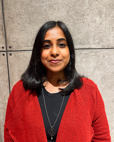
Dr. Anat Nitzan
Lab Manager


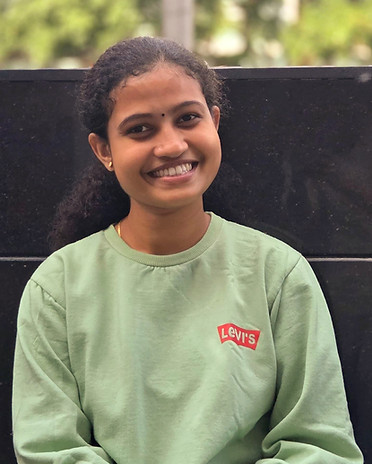
Anupreet Saini
PhD student